Bioimaging
Setup for live cell fluorescent imaging microscopy. This imaging setup shown was used for the online measurements of biochemical signaling in living cells stimulated using different approaches. It consists of a monochromator and a Spectra-X 7 channels LED light source that allows the selection of a specific wavelength and the quick shift between several wavelengths, a high sensitivity sCMOS camera, an inverted microscope with objectives equipped for high resolution fluorescence imaging, a perfusion chamber, a manifold that allows the quick and swift introduction of 8 different buffers to cells under a specific field and a data acquisition and analysis software.
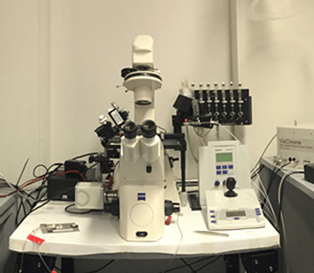
Live cell fluorescent imaging microscopy